Prof. Dr. Walter’s main interests lie in advancing bioimaging and biophotonics. His group aims at developing novel correlative microscopy solutions to facilitate imaging across scales in life sciences. His group combines advanced fluorescence microscopy with electron microscopy to visualize molecules within their subcellular context at high resolution using Correlative Light and Electron Microscopy (CLEM). Specifically, he envisions a unique advanced super-resolution and volume CLEM workflow under cryogenic conditions (Correlative Cryo-Super-Resolution Light and Focused Ion Beam Scanning Electron Microscopy) that pushes correlative microscopy further and allows his group – together with his many European collaboration partners – to tackle a variety of biomedical research questions that could not be answered before due to the lack of accessible solutions (ranging from nanoparticle-based cancer therapies to parasite infection of human cells).
His vision is to develop advanced multimodal imaging pipelines to zoom in from endogenous tissue or even living organisms to individual subcellular structures and relate their molecular dynamics to their overall anatomy.
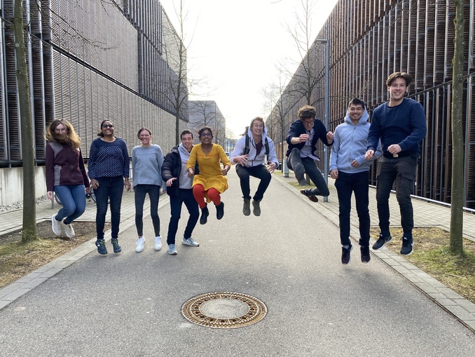
His team is a highly interdisciplinary group between physicis, engineering, optics and life sciences. There are several open positions available ranging from optical setups of home-built microscopes to automating image acquisition protocols for biological samples. Do not hesitate to contact andreas.walter@hs-aalen.de for further information or about available positions.
Aktuelle Mitarbeitende
- Research Associate: Michael Wagner

- Grant Manager COMULIS: Daniela Kuzdas-Wood
- PhD: Louisa Herbsleb
- Research Associate: Johannes Breitmeier
- Postdoctoral Scientist: Pavitra S. R.
- Lab technician/manager: Claudia Hintze
Research Associate: Leon Fuchs
- PhD: Michaela Salaková
BA-Student: Franziska Heeger
BA-Student: Jannick Heiberger
Ehemalige Mitarbeitende
- Robin Jürgens
- Clara Gündel
- Axel Wunderlich
Hansen, L., Heyer, W., Großbröhmer, C., Madesta, F., Sentker, T., Wang, J., Yuxi, Z., Zhang, H., Liu, M., Wang, J., Xi, Z., Yuhua, L., Liwen, W., Daniil, M., Nazim, H., Joel, H., Pekka, M., Yichao, Z., Zuopeng, T., Zhuoyuan, W., Yi, W., Hongchao, Z., Shunbo, H., Yi, Z., Qian, T., Lukas, F., Thomas, W., Bailiang, J., Christian, W., Jin, K., Dan, R., Marek, W., Tony C.W. M., Xi, J., Jinming, D., Mikael, B., Seyed-Ahmad, A., Yunzheng, Z., William, H., Tina, K., William, M. W., Alexandra, G., Aaron, C., Harrison, B., Yihao, L., Perrine, P.-G., Joakim, L., Nataša, S., Walter, A., Junyu, C., Reuben, D., Alessa, H., Mattias, P. ‘Learn2Reg 2024: New Benchmark Datasets Driving Progress on New Challenges. Machine Learning for Biomedical Imaging, 3, 775–791. https://doi.org/10.59275/j.melba.2025-gc8c (2025)
O’Connor, S., Rogers, D., Kobylynska, M., Geraets, J., Thaysen, K., Egebjerg, J. M., Brink, M. C., Herbsleb, L., Salakova, M., Fuchs, L., Alves, F., Feldmann, C., Ekman, A., Sheridan, P., Fyans, W., McEnroe, T., O’Reily, F., Fahy, K., Fleck, R. A., Wüstner, D., Simpson, J. C., Walter, A., Kapishnikov, S. ‘Demonstrating Soft X-Ray Tomography in the lab for correlative cryogenic biological imaging using X-rays and light microscopy.’ Sci Rep 15, 4549. https://doi.org/10.1038/s41598-025-29385-5 (2025)
Sokke Rudraiah, P., Herbsleb, L., Salakova, M., Gröger, H., Steyer, A. M., Alves, F., Feldmann, C., & Walter, A. Combining Correlative Cryogenic Fluorescence and Electron Microscopy and Correlative Cryogenic Super-Resolution Fluorescence and X-Ray Tomography-Novel Complementary 3D Cryo-Microscopy Across Scales to Reveal Nanoparticle Internalization Into Cancer Cells. Microscopy research and technique, 10.1002/jemt.70071. Advance online publication. https://doi.org/10.1002/jemt.70071 (2025)
Migasová, A., Zauška, Ľ., Zelenka, T., Volavka, D., Férová, M., Gulyásová, T., Tomková, S., Saláková, M., Kuchárová, V., Samuely, T., Bednarčík, J., Slabý, C., Máčajová, M., Bilčík, B., Schubert, T., Walter, A., Hornebecq, V., Huntošová, V., & Almáši, M. Histidine-modified UiO-66(Zr) nanoparticles as an effective pH-responsive carrier for 5-fluorouracil drug delivery system: A possible pathway to more effective brain cancer treatments. Chemical Engineering Journal. Advance online publication. https://doi.org/10.1016/j.cej.2025.167857 (2025)
Rudraiah PS, Camacho R, Fernandez-Rodriguez J, Fixler D, Grimm J, Gruber F, Kalaš M, Kremslehner C, Kuntner C, Kuzdas-Wood D, Lindblad J, Mannheim JG, Marchetti-Deschmann M, Avinoam O, Khan S, Paul-Gilloteaux P, Sampaio P, Sandbichler P, Sartori-Rupp A, Sladoje N, Verkade P, Walter A*, 'Correlated multimodal imaging in life sciences: lessons learnt'. Front. Biomater. Sci. 3:1338115. doi: 10.3389/fbiom.2024.1338115 (2025)
Nešić, N., Heiligenstein, X., Zopf, L., Blüml, V., Keuenhof, K. S., Wagner, M., Höög, J. L., Qi, H., Li, Z., Tsaramirsis, G., Peddie, C. J., Stojmenović, M., & Walter, A*. Automated segmentation of cell organelles in volume electron microscopy using deep learning. Microscopy Research and Technique, 87(8), 1718–1732. https://doi.org/10.1002/jemt.24548 (2024).
Bischof, J., Fletcher, G., Verkade, P. et al. Multimodal bioimaging across disciplines and scales: challenges, opportunities and breaking down barriers. npj Imaging 2, 5 (2024). https://doi.org/10.1038/s44303-024-00010-w (2024)
Bhattacharjee, P., Szabo, A., Dungel, P., Streli, C., Walter, A. Correlated Multimodal Imaging in Bone of Regeneration - A Showcase of Bisphosphonate-Treated Murine Jawbones. In: Walter, A., Slezak, P., Mueller, R., Kerckhofs, G., Bajoghli, B. (eds) Bioimaging in Tissue Engineering and Regeneration. Reference Series in Biomedical Engineering. Springer, Cham. (2024).
Praschl, C., Zopf, L.M., Kiemeyer, E., Langthallner, I., et int, Walter, A.. U-Net based vessel segmentation for murine brains with small micro-magnetic resonance imaging reference datasets. PLoS ONE 18(10): e0291946. https://doi.org/10.1371/journal.pone.0291946 (2023)
McLaren, M., Conners, R., Isupov, M.N., Gil-Diez, P., Gambell, L., Gold, V., Walter, A. et al. CryoEM reveals that ribosomes in microsporidian spores are locked in a dimeric hibernating state. Nat Microbiol 8, 1834–1845. https://doi.org/10.1038/s41564-023-01469-w (2023).
Cunha, F.F., Blüml, V., Zopf, L.M., Walter, A.*, et al., "Lossy Image Compression in a Preclinical Multomodal Imaging Study", J Digit Imaging https://doi.org/10.1007/s10278-023-00800-5 (2023)
Keuenhof, K.S., Kavirayani, A., Reier, S., Geyer, S.H., Weninger, W.J., Walter, A.*, ‘High-Resolution Episcopic Microscopy (HREM) in Multimodal Imaging Approaches.’ Biomedicines (2022) https://doi.org/10.3390/biomedicines9121918
Walter, A.*, Mannheim, J., Caruana, C., ‘Imaging Modalities in Biological and Preclinical Research: A Compendium.’ IoP-IPEM ebook series in Physics & Engineering in Medicine & Biology (2021) https://doi.org/10.1088/978-0-7503-3059-6– *Volume Editor
Walter, A.*, Paulino, C., Heuser, T., ‘Cryo-Electron Microscopy’ in ‘Imaging Modalities in Biological and Preclinical Research: A Compendium.’ IoP-IPEM ebook series in Physics & Engineering in Medicine & Biology (2021) https://doi.org/10.1088/978-0-7503-3059-6ch25
Walter, A.*, Stylianou, A., ‘Light Microscopy’ in ‘Imaging Modalities in Biological and Preclinical Research: A Compendium.’ IoP-IPEM ebook series in Physics & Engineering in Medicine & Biology (2021) https://doi.org/10.1088/978-0-7503-3059-6ch1
Walter, A.*, Pereiro, E., Harkiolaki, M., ‘Soft X-Ray Microscopy’ in ‘Imaging Modalities in Biological and Preclinical Research: A Compendium.’ IoP-IPEM ebook series in Physics & Engineering in Medicine & Biology (2021) https://doi.org/10.1088/978-0-7503-3059-6ch25
Verkade, P., Collinson, L., Munoz-Baruttia, A., Walter, A.*, ‘Emerging Imaging Technologies & Outlook’ in ‘Imaging Modalities in Biological and Preclinical Research: A Compendium.’ IoP-IPEM ebook series in Physics & Engineering in Medicine & Biology (2021) https://doi.org/10.1088/978-0-7503-3747-2ch31
Karreman, M., Pacureanu, A., Bosch Pinol, C., Schaefer, A., Walter, A.*, ‘Correlated Multimodality Imaging Across Scales’ in ‘Imaging Modalities in Biological and Preclinical Research: A Compendium.’ IoP-IPEM ebook series in Physics & Engineering in Medicine & Biology (2021) https://doi.org/10.1088/978-0-7503-3747-2ch23
Reier, S., Turyanskaya, A., Heimel, P., Frischauf, N., Meusburger, D., Heuser, T., Drexler, N., Cristina, S., Plochberger, B., Slezak, P., Dungel, P., Slezak, P., Szabo, A., Walter, A.*, ‘Cross-Modality Imaging of Bisphosphonate-Treated Murine Jawbones.’ Analyst, 146: 4683-4699 (2021) https://doi.org/10.1039/d0an02373f
Zopf, L., Jelena, Z., Mitterhauser, M., Helbich, T., Mengyang, L., Slezak, P., Weninger, W., Geyer, S., Kavirayani, A., Buehler, K., Walter, A.*, ‘Cross-Modality Imaging of Murine Tumor Vasculature – A Feasibility Study.’ Molecular Imaging & Biology (2021) https://doi.org/10.1007/s11307-021-01615-y
Keuenhofer, K., Macfelda, K., Streli, C., Schoefer, C., Weninger, W., Geyer, S., Slezak, P., Zopf, L., Wanek, T., Walter, A.*, ‘Multimodality imaging beyond CLEM: Showcases of combined in-vivo preclinical imaging and ex-vivo microscopy to detect murine mural vascular lesions.’ Methods in Cell Biology, 162:389-415 (2021) https://doi.org/10.1016/bs.mcb.2020.10.002
Walter, A., Verkade, P., ‘Community Efforts and Research Infrastructure for (Multimodality) BioImaging.’ Methods in Cell Biology, 162: 417-430 (2020) https://doi.org/10.1016/bs.mcb.2020.12.010
Walter, A.*, Paul-Gilloteaux, P., Plochberger, B., Verkade, P., Sefc, L., Mannheim, J., Unterhuber, A., Ogris, M., Marchetti-Deschmann, M., Handschuh, S., Dror, F., ‘Correlated Multimodality Imaging: Expanding the Biomedical Horizon.’ Front. Phys. 8:47 (2020) https://doi.org/10.3389/fphy.2020.00047
Roth, M., Gallaher, S., Westcott, D., Iwai, M., Louie, K., Mueller M., Walter, A., Foflonker, F., Bowen, B., Ataii, N., Song J., Chen J., Blaby-Haas, C., Larabell, C., Auer, M., Northern T., Merchant, S., Niyogi, K., ‘Regulation of Oxygenic Photosynthesis during Trophic Transitions in the Green Alga Chromochloris zofingiensis.’ Plant Cell, 31:579–60 (2019) https://doi.org/10.1105/tpc.18.00742
Roth, M., Cokus, J., Walter, A., Gallaher, S., Lopez, D., Erickson, E., Endelman, B., Westcott, D., Larabell, C., Merchant, S., Pellegrini, M., Niyogi, K., ‘Chromosome-level genome assembly and transcriptome of the green alga Chromochloris zofingiensis illuminates astaxanthin production.‘ Proc. Natl. Acad. Sci. USA (2017) https://doi.org/10.1073/pnas.1619928114
Walter, A., Steltenkamp, S., Schmitz, S., Holik, P., Sachser, R., Huth, M., Rhinow, D., Kühlbrandt, W., ‘Towards an optimum design for electrostatic phase plates.‘ Ultramicroscopy, 153:22-31 (2015) https://doi.org/10.1016/j.ultramic.2015.01.005
Gold, V., Raffaele, I., Walter, A., Pfanner, N., van der Laan, M., Kühlbrandt, W., ‘Visualizing active membrane protein complexes by electron cryotomography.‘ Nature Communications, 5:4129 (2014) https://doi.org/10.1038/ncomms5129
Walter, A., Chapuis, C., Huet, S., Ellenberg, J., ‘Crowded chromatin is not sufficient for heterochromatin formation and not required for its maintenance.‘ Journal of Structural Biology, 184:445-453 (2013) https://doi.org/10.1016/j.jsb.2013.10.004
Daum, B., Walter, A., Horst, A., Osiewacz, H., Kühlbrandt, W., ‘Age-dependent dissociation of ATP synthase dimers and loss of inner-membrane cristae in mitochondria.‘ Proc. Natl. Acad. Sci. USA, 110:15301-15306 (2013) https://doi.org/10.1073/pnas.1305462110
Walter, A., Muzik, H., Vieker, H., Turchanin, A., Beyer, A., Gölzhäuser, A., Lacher, M., Steltenkamp, S., Schmitz, S., Holik, P., Kühlbrandt, W., Rhinow, D., ‘Practical aspects of Boersch phase contrast electron microscopy of biological specimens.‘ Ultramicroscopy, 116:62-72 (2012) https://doi.org/10.1016/j.ultramic.2012.03.009
Barton, B., Rhinow, D., Walter, A., Schröder, R., Benner, G., Majorovits, E., Niebel, H., Müller, H., Haider, M., Lacher, M., Schmitz, S., Holik, P., Kühlbrandt, W., ‘In-focus electron microscopy of frozen-hydrated biological samples with a Boersch phase plate.‘ Ultramicroscopy, 111:1696-1705 (2011) https://doi.org/10.1016/j.ultramic.2011.09.007
Open positions of this working group and the ZOT can be found here.
Research projects of this working group and the ZOT can be found here.
COMULIS (Correlated Multimodal Imaging in Life Sciences) is an EU-funded COST Action that aims at fueling urgently needed collaborations in the field of correlated multimodal imaging (CMI), promoting and disseminating its benefits through showcase pipelines, and paving the way for its technological advancement and implementation as a versatile tool in biological and preclinical research. CMI combines two or more imaging modalities to gather information about the same specimen. It creates a composite view of the sample with multidimensional information about its macro-, meso- and microscopic structure, dynamics, function and chemical composition. Since no single imaging technique can reveal all these details, CMI is the only way to understand biomedical processes and diseases mechanistically and holistically. CMI relies on the joint multidisciplinary expertise from biologists, physicists, chemists, clinicians and computer scientists, and depends on coordinated activities and knowledge transfer between academia and industry, and instrument developers and users. Due to its inherently multidisciplinary and crossfunctional nature, an interdisciplinary network such as this Action is indispensable for the success of CMI. Nevertheless, there is currently no European network in the field. Existing scattered efforts focus on correlated light and electron microscopy or (pre)clinical hybrid imaging. This Action will consolidate these efforts, establish commonly-accepted protocols and quality standards for existing CMI approaches, identify and showcase novel CMI pipelines, bridge the gap between preclinical and biological imaging, and foster correlation software through networking, workshops and open databases. The network will raise awareness for CMI, train researchers in multimodal approaches, and work towards a scientific mindset that is enthusiastic about interdisciplinary imaging approaches in life sciences.
The COMULIS network has now been extended and funded by the Chan Zuckerberg Initiative (COMULISglobe) to establish a global community, ensure long-term funding, and include clinical diagnostics in the multimodal imaging efforts and showcases.