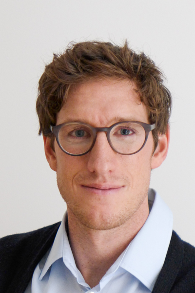
Studienprojekte und eine Zusammenfassung der Forschungsaktivitäten von Prof. Dr. Walter am Zentrum für optische Technologien (ZOT) und innerhalb der COST Action COMULIS finden Sie auf seiner ZOT-Seite.
The great strength of fluorescence microscopy (FM) at room temperature is its ability to study living cells. In cases where the dynamics are too fast and the sample needs to be immobilized for imaging (e.g., for super-resolution (SR) FM), chemical fixation is usually the method of choice. However, chemical fixation is associated with structural changes in the sample and can lead to artifacts in the microscopy images, which in turn can lead to misinterpretation of biological functions and mechanisms. Rapid freezing (vitrification) of the sample allows immobilization in a near-natural state. Structural preservation is particularly important for microscopy techniques that achieve very high resolution. Therefore, the main application of cryo-FM is in combination with cryo-electron microscopy (EM). Nevertheless, cryo-FM can also be combined with SR imaging and serve as an alternative to classical SR-FM in chemically fixed samples. In this project, we aim to develop a unique cryogenic SR-FM below the Abbe diffraction limit with structured illumination (SIM). The cryogenic SIM setup will visualize molecules with twice the lateral resolution compared to conventional FM and enable optical sections. This setup sets the stage for correlating FM with cryo-EM (in particular focused ion beam scanning electron microscopy) and addressing clinically relevant research questions. The correlation of microscopy techniques under cryogenic conditions is important because it directly images specific molecules in their original cellular context: While EM allows visualization of the subcellular architecture of a cell, molecular complexes within that cell can only be uniquely localized by FM.
This project will consolidate and extend a collaborative and innovative network promoting MultiModal Imaging and analysis across scales (MMI) from biological research to clinical diagnostics, and establish a global multimodal imaging association (COMULISglobe) to ensure long term sustainability. MMI integrates the best features of combined techniques and overcomes limitations faced when applying single modalities independently. MMI relies on the joint expertise from biologists, physicists, chemists, clinicians, and computer scientists, and depends on coordinated activities and knowledge transfer between technology developers and users. To achieve this inherently interdisciplinary goal, it is indispensable to establish a network of scientists across continents and disciplines, from academia and industry, including transnational research facilities (e.g. synchrotrons, Euro-BioImaging ERIC), to foster and market MMI as a versatile tool in biomedical research and diagnostics. We will capitalize on COMULIS, a European initiative (comulis.eu), and extend it globally and sustainably. The network will raise awareness of the manifold benefits of MMI, train researchers, and promote a scientific mindset enthusiastic about interdisciplinary imaging and analysis. The MMI network will help bridge the gap between biological and clinical imaging, identify, fund, and showcase novel multimodal pipelines, and develop, evaluate, and publish correlation software through dedicated networking activities, including conferences, training schools, open databases, and fellowships for lab exchanges, access to research infrastructures, and conference attendance. All outputs of the project will be open access.
CLEXM is a Marie Sklodowska-Curie Doctoral Networks Action (MSCA-DN), funded by the European Union under Horizon Europe. The objective of MSCA-DN Project is to implement doctoral programmes by partnerships of organisations as well as to train highly skilled doctoral candidates, stimulate their creativity, enhance their innovation capacities and boost their employability in the long-term. Partner organisations include the Institut Pasteur in Paris, University College Dublin, and ALBA synchrotron in Barcelona. CLEXM addresses an urgent need for collaborations in the field of correlative multimodal imaging. It will gather and integrate information from complementary imaging modalities to create a more complete view of biomedical processes of disease and drug therapy research.
Bei der Krebstherapie durch Strahlen- und Chemotherapie werden auch gesunde Zellen beschädigt. Mit einer neuen Tumorbekämpfung möchte das interdisziplinäre Verbundprojekt „NanoLYRIC“ der Hochschule Aalen, des Karlsruher Instituts für Technologie (KIT) und der Universitätsmedizin Göttingen (UMG) gezielt und effizienter als bisher Krebszellen abtöten und Nebenwirkungen auf das gesunde Gewebe reduzieren. Dazu werden am KIT in der Arbeitsgruppe von Prof. C. Feldmann neuartige Nanopartikel entwickelt, die mit Chemotherapeutika beladen sind und Gold als schweres Element enthalten. Diese nehmen bei der Strahlentherapie verstärkt Strahlung in das Tumorgewebe auf und setzen die Chemotherapeutika erst unter Bestrahlung selektiv und nur im Tumor frei. Diese gleichzeitige, durch die Nanopartikel vermittelte Kombination von Strahlen- und Chemotherapie verstärkt die Wirkung der Krebstherapie durch selektives Abtöten nur der Krebszellen und Verschonung von gesundem Gewebe.
Diese neue nanopartikelbasierte Therapie wird von der UMG von Prof. F. Alves, Prof. C. Dullin und Prof. S .Rieken auf ihre Anreicherung im Tumor und ihre Wirkung präklinisch evaluiert. Für die Auswertung werden von der Hochschule Aalen von Prof. A. Walter und Prof. C. Neusüß und der UMG skalenübergreifende bildgebende Verfahren verwendet.
Ziel ist es, die Nebenwirkungen der Therapie auf das gesunde Gewebe bzw. den Gesamtorganismus zu reduzieren und gleichzeitig die therapeutische Wirksamkeit zu verbessern und somit die Lebensqualität und Überlebenschancen von Patient:innen zu erhöhen.

Prof. Dr. Andreas Walter
Sprechzeiten
zu festen Zeiten| Mittwoch | 14:00 - 14:30 G1 2.11 |
